فئات
الأشعة الطبية
معالجات الأفلام الآلية
معالجات الأفلام اليدوية
تصنيف: أفلام الأشعة السينية
كاسيت الأشعة السينية
تكثيف الشاشات
أضواء الظلام
حلول تطوير الأفلام
فيلم مجفف
مشاهدي الأفلام
الشماعات الفيلم
علامات الفيلم
صناديق نقل الفيلم
صناديق تخزين الأفلام
المدرجات الصدر
أجهزة قياس الجرعات بالأشعة السينية
جداول الأشعة السينية
غرفة الأشعة السينية
مولد الأشعة السينية
التصوير بالرنين المغناطيسي المعدات غير المغناطيسية
منتجات جديدة


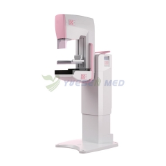
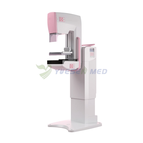


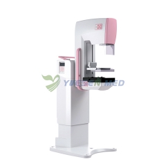


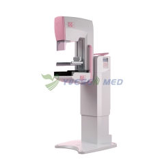


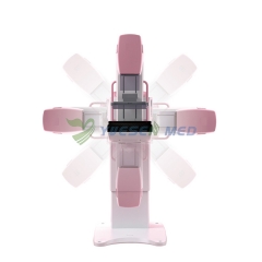


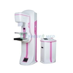
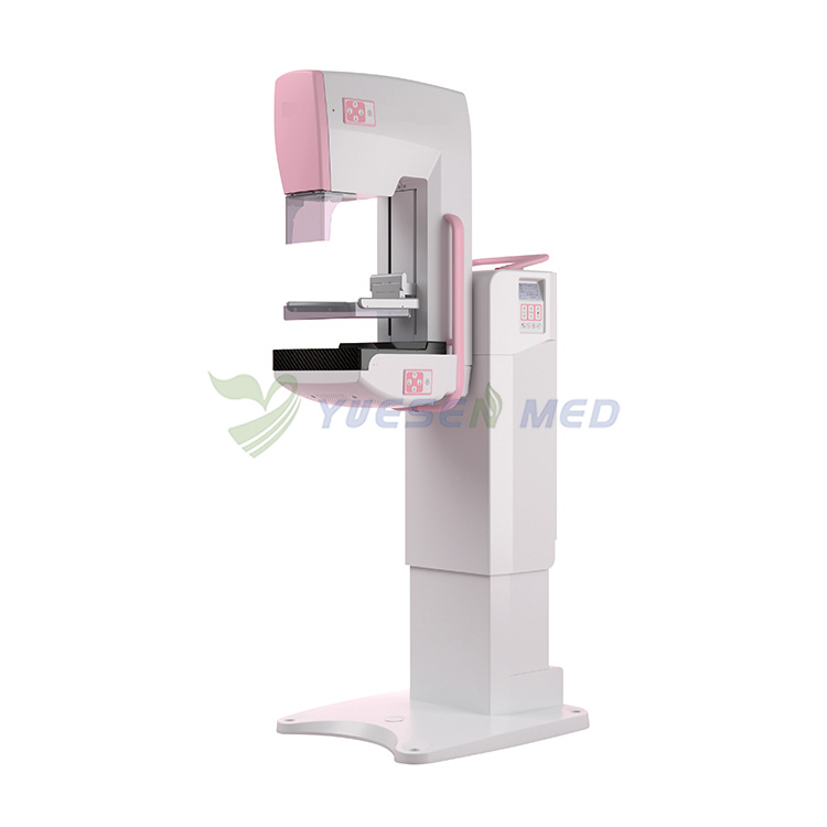
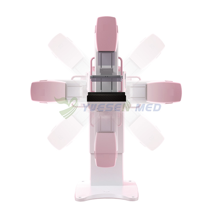
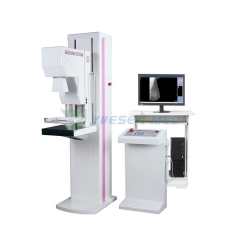
آلة تصوير الثدي بالأشعة السينية المتنقلة
رقم البند.: YSX-DM300
أحدث آلة تصوير الثدي بالأشعة السينية المتنقلة للبيع بسعر جيد.
| معلمات المنتج | |
| ماركة: | YSENMED |
| MOQ: | 1 |
| حزمة: | عبوة خشبية |
| الشحن من: | Guangzhou |
| المهلة: | 1-3 أيام |
| التخصيص: | Yes |
وصف
آلة تصوير الثدي بالأشعة السينية المتنقلة
| Item | Description | |
| 1 | FULL SYSTEM | |
Optional:Tungsten Anode, Focal Spot Size: 0.1 mm (Small)/ 0.3 mm (Large)
|
||
| 2 | High-Voltage Generator | |
| a) Frequency:≥100KHz; b) Tube voltage Range: 20 kV ~ 40 kV in 1 kV step; c) Max Output: 4 kW; d) Max tube voltage:40kV; e) Max tube current: 140mA; f) Max current time product: ≥630mAs; g) Ripple: <4%. |
||
| 3 | X-Ray Tube | |
| TUBE | Standard:
|
|
Optional:
|
||
| 4 | Beam Limiting Device | |
|
||
| 5 | C-arm Assembly |
|
|
||
| 6 | Compression Device | |
|
||
| 7 | Local Operation and Display | |
|
||
| 8 |
|
|
|
||
| 9 | Digital Image Detector | |
|
||
| 10 | Acquisition Station | |
|
||
| 11 | Automatic Exposure Control | |
|
||
| 12 | Manuals | |
|
||

YOUR MAY ALSO LIKE ...
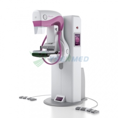
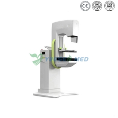
نظام تصوير الثدي الرقمي ثلاثي الأبعاد بتقنية التصوير المقطعي للثدي YSX-DM300A