فئات
الأشعة الطبية
معالجات الأفلام الآلية
معالجات الأفلام اليدوية
تصنيف: أفلام الأشعة السينية
كاسيت الأشعة السينية
تكثيف الشاشات
أضواء الظلام
حلول تطوير الأفلام
فيلم مجفف
مشاهدي الأفلام
الشماعات الفيلم
علامات الفيلم
صناديق نقل الفيلم
صناديق تخزين الأفلام
المدرجات الصدر
أجهزة قياس الجرعات بالأشعة السينية
جداول الأشعة السينية
غرفة الأشعة السينية
مولد الأشعة السينية
التصوير بالرنين المغناطيسي المعدات غير المغناطيسية
منتجات جديدة


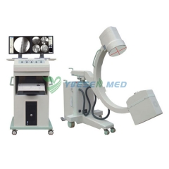
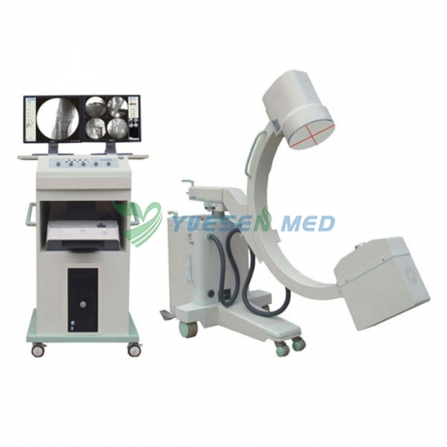
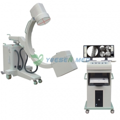


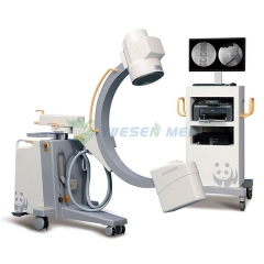
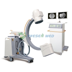
3.5KW/5KW Mega-pixel الرقمية C-arm YSX-C35D
رقم البند.: YSX-C35D
عالية الأداء 3.5KW 5KW جهاز الأشعة السينية الطبية الرقمية C-arm YSX-C35D انخفاض السعر
| معلمات المنتج | |
| ماركة: | YSENMED |
| MOQ: | 1 |
| حزمة: | عبوة خشبية |
| الشحن من: | Guangzhou |
| المهلة: | 1-3 أيام |
| التخصيص: | Yes |
وصف
High Performance 3.5KW 5KW Mega-pixel Digital C-arm X-ray Machine YSX-C35D


1.Combined X-ray tube & generator
2.9inch Toshiba Image Intensifier
3.get the clearest images with minimum dose
YSX-C35D Technical parameters and configuration:
| Main components | Technique parameter | |||
| IMD X-ray tube | Model:YSX-C35D | Model:YSX-C50D | ||
| Power:3.5kW | Power:5.0kW | |||
| Double focus fixed anode focus:1.5mm / 0.6mm | Double focus rotational anode focus: 0.3mm / 0.6mm | |||
| Anode Heat Capacity:40kHu X-ray tube sleeve heat capacity:667kHu | Anode Heat Capacity:200kHu X-ray tube sleeve heat capacity:800kHu | |||
| High Voltage X-ray Generator | Working frequency:40kHz | |||
| Image intensifier | Toshiba 9"(including 3 fields of view 4.5"/ 6"/9") | |||
| Tube voltage | Fluoroscopy Radiograph:40kV-110kV | Fluoroscopy Radiograph:40kV-120kV | ||
| Tube current | Fluoroscopy: 0.5-4mA Enhanced Automatic Fluoroscopy: 1mA-8mA Pulse Fluoroscopy :2pps;8mA | |||
| mAs | 1mA.s-250mA.S | |||
| Fluoroscopy mode | Manual Fluoroscopy; Semi-Automatic Fluoroscopy;Automatic Fluoroscopy; Enhanced Automatic Fluoroscopy; Pulse Fluoroscopy | |||
| Intelligent exposure control | No matter the object is in the center or on the fringing field of the image intensifier, the images are the same. The exposure dose is reduced. | |||
| Iris beam limiting device | when not exposure, keep in the state of Iris Beam limiting preview ,when exposure,keep blanking cycle track | |||
| General radiography | 40kV-120kV; 1.0mA.s-250mA.s | step length 1 kV; 1.0mA.s | ||
| Digital radiography (digital spot film) | 40kV-120kV; Max 16mA | Common screen film radiography is eliminated. Digital radiography can get better images | ||
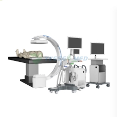
| C-arm machinery index | Distance between focus and window≧900mm, arc depth≧650mm, horizontal moving range: 200mm vertical moving range: 400mm | |||
| Tilt angle: ±12.5° rotation: ±180°corner: 125°(-35°-- +90°) | ||||
| Laser Orientation | Laser positioning function can help to position precisely and reduce exposure times in the operation, reduce unnecessary radiation. | |||
| Radiography mode | ||||
| Image intensifier & Real-time digital image(1.3 million pixel) processing system | ||||
| (CCD)Digital video camera | million pixels high-definition, high-speed CCD camera, a 1024 × 1024 matrix, 30 frames per second image acquisition, through real-time adjustment of window width, window level, noise reduction, sharpening and Gain adjustment to ensure high-quality and high-resolution images. | |||
| Image resolution index | Grey level:10 Resolution:22 lp/cm | |||
| Camera virtual rotation design | No X-ray needed, also called digital image rotation,display the right angle when exposure, superior to camera mechanical rotation | |||
| Image processing system | 1.Real time edge enhancement(sharpening) | Image sharpening is also called edge enhancement, that is, to make blurry edge of the image clearer | ||
| 2.Real time adjustment of the window width and window level | Obtain images by real-time adjustment of window width,window level, displaying images with different brightness. | |||
| 3.Real time(static)multiform noise reduction | Noise reduction, and improve image clarity | |||
| 4.Real tim GAIN adjustment | Can amplify image signal, reduce x-ray dose, suitable for patient with super large size. | |||
| 5.Real tim dynamic brightness compensation and R calibration functions | Through dynamic logarithmic transformation, linear reduction the system signals and r calibration technology, dark parts of the image can be enhanced and clear rich-layer images can be obtained | |||
| 6.Image conversing ,up-down and right-left rotating, and frame freezing | ||||
| 7.Patient information management, Clinic report and print | ||||
| 8.The Image can be storage as JPG and DICOM 3.0 format, can be linked to hard-disk, USB and DVD and printer, meet the need of sharing the network information, communication and storage in hospital | ||||
| Display | Display2 17"TFT-LCD display | |||
| Printer | Printer Laser / jet Printer | |||
| Standard configuration | 1.Combined tube(5.0kW)IMD | 1 set; | ||
| 2.Image intensifier (9 inch 3 fields of view) Toshiba | 1 set | |||
| 3.C-arm stander (with full C-arm balance system) | 1 set; | |||
| 4.Exported grid | 1 set | |||
| 5.Laser positioning | 1 set; | |||
| 6.1.3million pixel 1024×1280×10bit camera | 1 set; | |||
| 7.Electric adjustable beam limiting device | 1 set | |||
| 8.17 inch LCD high resolution screen | 2 sets | |||
| 9.Real-time digital imageprocession system (station) | 1 set; | |||
| 10.Printer | 1 set | |||

YOUR MAY ALSO LIKE ...
كاشف اللوحة المسطحة 15 كيلو وات، نظام الأشعة السينية الرقمي ذو الذراع C YSX-C715