


catégories
Radiologie médicale
Processeurs de film automatique
Processeurs manuels de film
X-ray films
Cassettes à rayons X
écrans renforçateurs
Feux De Chambre Noire
Film Développement De Solutions
Séchage Machines Film
Les Téléspectateurs De Films
Cintres De Film
Marqueurs Film
Des Boîtes De Transfert Du Film
Film stocker des boîtes
Stands De La Poitrine
Les Dosimètres à Rayons X
Tables De Rayons X
X-ray Chambre
Générateur Radiologique
Matériel Amagnétique IRM
Produits chauds
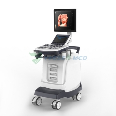


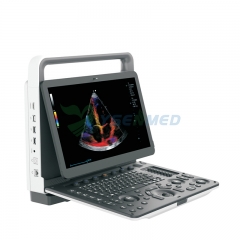
Système De Echographie Doppler Couleur YSB-L5
No. d'article: YSB-L5
Système De Echographie Doppler Couleur YSB-L5 Prix / Ultrason portatif de Doppler de couleur à vendre de Chine.
Description
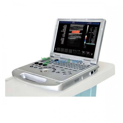
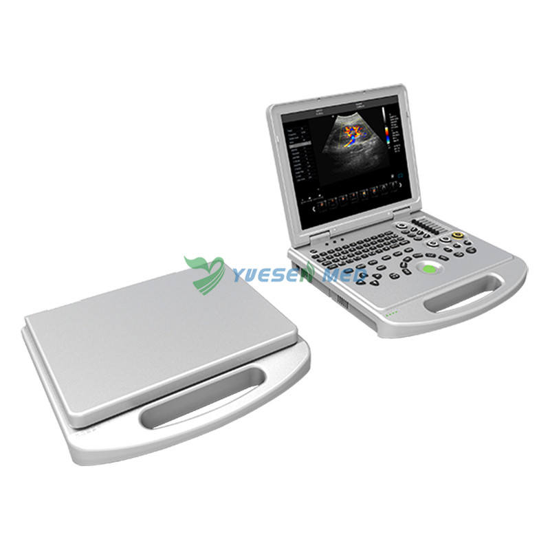
Portable color doppler ultrasound YSB-L5

Parameter:
Detection depth: ≥ 300mm.
Lateral resolution: ≤ 1mm (depth ≤ 80mm)
≤ 2mm (80
≤2mm(80
Precision geometric position: Horizontal ≤ 10%.
Longitudinal ≤ 10%.
Signal channels: 64 channels
Display: 15 inch LED monitor.
PAL output interface
Grey Scale: 256
Power range: AC220V ± 10% 3A.
Host Power: DC12.8V 11.5A
Input power: ≤ 300VA.
Continuous working time: ≥ 8h.
-Application
Abdomen, OB&GYN, cardiology, vascular and small parts, urology, musculoskeletal, pediatrics and etc
-Displaying mode
B, 2B, 4B, left&right, B|M, B|D, PW, M, B mode, part zoom, B|C|D, B|C|M, B|C,
duplex, PW, CFM, CPA
-Signal processing
Full-digital beam forming,
dynamic filter, orthogonal
demodulating,
space-time filter, dynamic
real-time receiving focusing, RDA, DRA, spectral processing, CFM processing
-Image processing
THI, speckle-reduction, color coder, frame averaging, micro- angle adjustment, wall filter, 256
grey scale, scanning angle/width control, composit processing of tissue and blood flow image
-General measurement
B mode: distance, angle, perimeter and area (ellipse method, Trace method), volume, histogram,
cross-section diagram
M mode: cardiac rate, time, distance, speed.
-Measurement & report packages
GYN(four edition for GA calculation), cardiac, vascular, urology, andriatrics, peripheral vascular, multiple births, orthopedic surgery and etc.
-Storage function
Probe parameter, image, cine loop, measurement data and report
-Cine loop
Operated by automatically and manually, speed optional, searching cine loop, forward/ backward cine loop
- Input/output interface
VGA, network, USB, VIDEO, parallet communication port, serial communication port
-Standard configuration
Main unit, 3.5MHz Multi-frequency convex probe(2.0, 3.0, 3.5, 4.0,5.5MHz),
15" LED monitor, USB port
DICOM3.0
-Optional configuration
Probe:
| High frequency linear probe | 128 elements R40 wideband | Multi-frequency 6.0,6.5,7.5,10,12MHz |
| Trans-vaginal probe | 128 elements R10 wideband | Multi-frequency 5.0,6.0,6.5,7.5,9.0MHZ |
| Phased array probe | 64 elements R20 wideband | Multi-frequency 2.0,2.1,3.5,4.0,5.0 MHz |

YOUR MAY ALSO LIKE ...
Full Digital Color Doppler Ultrasonic Diagnostic System YSB-VIV20
Scanner portatif rentable d'échographie Doppler couleur 4D YSB-M70

