Hot Products
YSX500D 50kW DR system set up and put into service in Cambodia.
YSENMED YSX500D 50kW digital x-ray system has been successfully set up and put into service in a hospital in Cambodia.
YSX056-PE serving as a vehicle-mounted x-ray in the Philippines
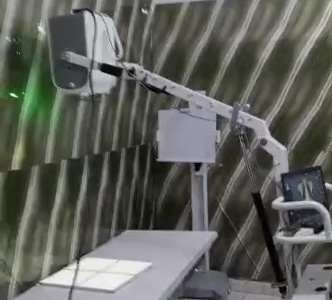
YSX056-PE 5.6kW portable x-ray unit has been adapted to fit on a truck, to provide mobile x-ray examination service for remote communities in the Philippines.
X Ray Machine To Zimbabwe
x ray machine, 50KW x ray machine
Microscope To Malawi
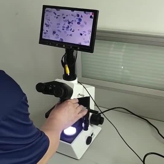
Achromatic objectives: 4X、10X、40X(S), 100X(S、Oil) Wide field eyepiece: WF10X(WF16X for option) Eyepiece head: Sliding binocular head inclined at 45° Stage: Double layer mechanical stage size 140X140mm, moving range 75X45mm Focusing: Coaxial coarse and
The Visionary Tool: Exploring the Eye Surgical Operating Microscope
Views : 1863
Update time : 2024-04-30 12:04:00
Introduction
In the world of modern medicine, advancements in technology have revolutionized the way surgeries are performed, especially in delicate procedures like eye surgery. One such remarkable innovation is the eye surgical operating microscope. This sophisticated device has become an indispensable tool for ophthalmologists, enabling them to perform intricate procedures with unparalleled precision and accuracy. In this article, we delve into the intricacies of the eye surgical operating microscope, its components, functionalities, and the invaluable role it plays in ensuring successful eye surgeries.

Understanding the Eye Surgical Operating Microscope
The eye surgical operating microscope, also known as an ophthalmic microscope, is a specialized optical instrument designed for performing surgeries on the delicate structures of the eye. It consists of a high-powered microscope mounted on a stable floor stand or ceiling mount, providing the surgeon with a magnified and illuminated view of the eye's internal structures.
Components of an Eye Surgical Operating Microscope
1. Binocular Microscope Head: At the heart of the eye surgical operating microscope is the binocular microscope head, equipped with adjustable eyepieces and objective lenses. This component allows the surgeon to view the surgical field in three-dimensional detail and with enhanced clarity.
2. Light Source: A powerful light source, typically an LED or xenon lamp, is integrated into the microscope to illuminate the surgical area. The intensity and angle of the light can be adjusted to provide optimal illumination without causing glare or discomfort to the patient.
3. Magnification System: The magnification system of the microscope consists of interchangeable objective lenses, allowing the surgeon to adjust the magnification level according to the specific requirements of the procedure. High magnification levels enable precise visualization of minute structures within the eye.
4. Fine Focus Mechanism: A fine focus mechanism allows the surgeon to make minute adjustments to the focus of the microscope, ensuring sharp and clear visualization throughout the surgery. This feature is crucial for maintaining accuracy during delicate maneuvers.
Functionalities of the Eye Surgical Operating Microscope
1. Enhanced Visualization: The primary function of the eye surgical operating microscope is to provide the surgeon with a clear and magnified view of the surgical site. This enhanced visualization is essential for identifying anatomical structures, navigating intricate pathways, and performing precise surgical maneuvers.
2. Illumination Control: The microscope's adjustable light source allows the surgeon to control the intensity, direction, and angle of illumination, minimizing shadows and glare that may obscure the surgical field. This precise control ensures optimal visibility and enhances surgical precision.
3. Integrated Imaging Systems: Many modern eye surgical operating microscopes are equipped with integrated imaging systems, such as digital cameras and video recording capabilities. These systems enable the surgeon to capture high-resolution images and videos of the surgical procedure for documentation, analysis, and education purposes.
Applications of the Eye Surgical Operating Microscope
1. Cataract Surgery: The eye surgical operating microscope plays a crucial role in cataract surgery, allowing the surgeon to visualize and remove the cloudy lens with unparalleled precision. High magnification and illumination enable the precise placement of intraocular lens implants, ensuring optimal visual outcomes for patients.
2. Retinal Surgery: In retinal surgery, where delicate maneuvers are required to repair retinal detachments or remove epiretinal membranes, the eye surgical operating microscope provides the surgeon with a clear and magnified view of the retina and vitreous cavity. This enables precise tissue dissection and membrane peeling, minimizing the risk of complications.
3. Corneal Transplantation: During corneal transplantation procedures, such as penetrating keratoplasty or Descemet's membrane endothelial keratoplasty (DMEK), the eye surgical operating microscope facilitates the precise placement and suturing of donor corneal tissue. High magnification and illumination are essential for achieving optimal graft alignment and stability.
Benefits of the Eye Surgical Operating Microscope
1. Precision and Accuracy: By providing magnified and illuminated views of the surgical field, the eye surgical operating microscope enables surgeons to perform complex procedures with unprecedented precision and accuracy.
2. Improved Surgical Outcomes: The enhanced visualization and control offered by the microscope result in improved surgical outcomes, reduced complication rates, and faster patient recovery times.
3. Educational Value: Eye surgical operating microscopes equipped with imaging systems allow surgeons to document and review surgical procedures, facilitating education, training, and knowledge sharing within the medical community.
Conclusion
The eye surgical operating microscope represents a pinnacle of technological innovation in the field of ophthalmic surgery. Its advanced optics, precise control features, and integrated imaging systems empower surgeons to perform complex procedures with confidence and proficiency. As technology continues to evolve, the eye surgical operating microscope will undoubtedly remain an indispensable tool in the pursuit of optimal eye health and visual outcomes for patients worldwide.
How does an eye surgical operating microscope work?
The eye surgical operating microscope works by magnifying and illuminating the surgical field, allowing the surgeon to visualize the delicate structures of the eye with enhanced clarity and precision. It consists of a binocular microscope head, a light source, interchangeable objective lenses, and a fine focus mechanism.
What are the main components of an eye surgical operating microscope?
The main components of an eye surgical operating microscope include the binocular microscope head, the light source, the magnification system with interchangeable objective lenses, and the fine focus mechanism. These components work together to provide the surgeon with a clear and magnified view of the surgical site.
What types of eye surgeries can be performed using an eye surgical operating microscope?
An eye surgical operating microscope is commonly used in various eye surgeries, including cataract surgery, retinal surgery, corneal transplantation, glaucoma surgery, and oculoplastic surgery. Its advanced optics and precise control features make it suitable for performing intricate procedures on the delicate structures of the eye.
How does the eye surgical operating microscope benefit surgeons and patients?
The eye surgical operating microscope benefits surgeons by providing them with enhanced visualization and control during surgical procedures, resulting in improved surgical outcomes and reduced complication rates. For patients, this means faster recovery times, fewer postoperative complications, and better visual outcomes.
Is training required to use an eye surgical operating microscope?
Yes, training is required to use an eye surgical operating microscope effectively. Surgeons undergo specialized training and hands-on practice to familiarize themselves with the microscope's functionalities, ergonomics, and operating techniques. This ensures safe and proficient use of the microscope during surgical procedures.
In the world of modern medicine, advancements in technology have revolutionized the way surgeries are performed, especially in delicate procedures like eye surgery. One such remarkable innovation is the eye surgical operating microscope. This sophisticated device has become an indispensable tool for ophthalmologists, enabling them to perform intricate procedures with unparalleled precision and accuracy. In this article, we delve into the intricacies of the eye surgical operating microscope, its components, functionalities, and the invaluable role it plays in ensuring successful eye surgeries.

Understanding the Eye Surgical Operating Microscope
The eye surgical operating microscope, also known as an ophthalmic microscope, is a specialized optical instrument designed for performing surgeries on the delicate structures of the eye. It consists of a high-powered microscope mounted on a stable floor stand or ceiling mount, providing the surgeon with a magnified and illuminated view of the eye's internal structures.
Components of an Eye Surgical Operating Microscope
1. Binocular Microscope Head: At the heart of the eye surgical operating microscope is the binocular microscope head, equipped with adjustable eyepieces and objective lenses. This component allows the surgeon to view the surgical field in three-dimensional detail and with enhanced clarity.
2. Light Source: A powerful light source, typically an LED or xenon lamp, is integrated into the microscope to illuminate the surgical area. The intensity and angle of the light can be adjusted to provide optimal illumination without causing glare or discomfort to the patient.
3. Magnification System: The magnification system of the microscope consists of interchangeable objective lenses, allowing the surgeon to adjust the magnification level according to the specific requirements of the procedure. High magnification levels enable precise visualization of minute structures within the eye.
4. Fine Focus Mechanism: A fine focus mechanism allows the surgeon to make minute adjustments to the focus of the microscope, ensuring sharp and clear visualization throughout the surgery. This feature is crucial for maintaining accuracy during delicate maneuvers.
Functionalities of the Eye Surgical Operating Microscope
1. Enhanced Visualization: The primary function of the eye surgical operating microscope is to provide the surgeon with a clear and magnified view of the surgical site. This enhanced visualization is essential for identifying anatomical structures, navigating intricate pathways, and performing precise surgical maneuvers.
2. Illumination Control: The microscope's adjustable light source allows the surgeon to control the intensity, direction, and angle of illumination, minimizing shadows and glare that may obscure the surgical field. This precise control ensures optimal visibility and enhances surgical precision.
3. Integrated Imaging Systems: Many modern eye surgical operating microscopes are equipped with integrated imaging systems, such as digital cameras and video recording capabilities. These systems enable the surgeon to capture high-resolution images and videos of the surgical procedure for documentation, analysis, and education purposes.
Applications of the Eye Surgical Operating Microscope
1. Cataract Surgery: The eye surgical operating microscope plays a crucial role in cataract surgery, allowing the surgeon to visualize and remove the cloudy lens with unparalleled precision. High magnification and illumination enable the precise placement of intraocular lens implants, ensuring optimal visual outcomes for patients.
2. Retinal Surgery: In retinal surgery, where delicate maneuvers are required to repair retinal detachments or remove epiretinal membranes, the eye surgical operating microscope provides the surgeon with a clear and magnified view of the retina and vitreous cavity. This enables precise tissue dissection and membrane peeling, minimizing the risk of complications.
3. Corneal Transplantation: During corneal transplantation procedures, such as penetrating keratoplasty or Descemet's membrane endothelial keratoplasty (DMEK), the eye surgical operating microscope facilitates the precise placement and suturing of donor corneal tissue. High magnification and illumination are essential for achieving optimal graft alignment and stability.
Benefits of the Eye Surgical Operating Microscope
1. Precision and Accuracy: By providing magnified and illuminated views of the surgical field, the eye surgical operating microscope enables surgeons to perform complex procedures with unprecedented precision and accuracy.
2. Improved Surgical Outcomes: The enhanced visualization and control offered by the microscope result in improved surgical outcomes, reduced complication rates, and faster patient recovery times.
3. Educational Value: Eye surgical operating microscopes equipped with imaging systems allow surgeons to document and review surgical procedures, facilitating education, training, and knowledge sharing within the medical community.
Conclusion
The eye surgical operating microscope represents a pinnacle of technological innovation in the field of ophthalmic surgery. Its advanced optics, precise control features, and integrated imaging systems empower surgeons to perform complex procedures with confidence and proficiency. As technology continues to evolve, the eye surgical operating microscope will undoubtedly remain an indispensable tool in the pursuit of optimal eye health and visual outcomes for patients worldwide.
How does an eye surgical operating microscope work?
The eye surgical operating microscope works by magnifying and illuminating the surgical field, allowing the surgeon to visualize the delicate structures of the eye with enhanced clarity and precision. It consists of a binocular microscope head, a light source, interchangeable objective lenses, and a fine focus mechanism.
What are the main components of an eye surgical operating microscope?
The main components of an eye surgical operating microscope include the binocular microscope head, the light source, the magnification system with interchangeable objective lenses, and the fine focus mechanism. These components work together to provide the surgeon with a clear and magnified view of the surgical site.
What types of eye surgeries can be performed using an eye surgical operating microscope?
An eye surgical operating microscope is commonly used in various eye surgeries, including cataract surgery, retinal surgery, corneal transplantation, glaucoma surgery, and oculoplastic surgery. Its advanced optics and precise control features make it suitable for performing intricate procedures on the delicate structures of the eye.
How does the eye surgical operating microscope benefit surgeons and patients?
The eye surgical operating microscope benefits surgeons by providing them with enhanced visualization and control during surgical procedures, resulting in improved surgical outcomes and reduced complication rates. For patients, this means faster recovery times, fewer postoperative complications, and better visual outcomes.
Is training required to use an eye surgical operating microscope?
Yes, training is required to use an eye surgical operating microscope effectively. Surgeons undergo specialized training and hands-on practice to familiarize themselves with the microscope's functionalities, ergonomics, and operating techniques. This ensures safe and proficient use of the microscope during surgical procedures.
Related News
Read More >>
 Introduction video of YSENMED YSXWJ-BX301B Digital Biological Microscope.
Apr .01.2025
Here we share the introduction video of YSENMED YSXWJ-BX301B Digital Biological Microscope.
 YSDR-VET320 veterinary DR & YSB-DU10V color ultrasound serving well in Kiram Veterinary Surgery in Uganda
Mar .31.2025
Kiram Veterinary Surgery in Uganda shares the clear images they have got from YSDR-VET320 veterinary digital x-ray system and YSB-DU10V vet color ultrasound machine.
 YSENMED YSFPD-R1012C mammo flat panel detector introduced and set up in a hospital in Senegal
Mar .30.2025
Senegalese hospital has upgraded its analog mammography x-ray system with YSFPD-R1012C mammo flat panel detector, and the images come out clear.
 YSX-mDR5A 5.3kW mobile DR system working fine in a clinic in Gambia
Mar .29.2025
YSENMED YSX-mDR5A 5.3kW mobile digital x-ray unit is working well in a clinic in Gambia.


