Categories
Medical Radiology
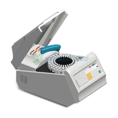
Auto film processors
Manual film processors
X-ray films
X-ray Cassettes
Intensifying screens
Darkroom lights
Film developing solutions
Film dryer
Film viewers
Film hangers
Film markers
Film transfer boxes
Film storing boxes
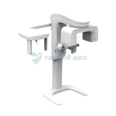
Chest stands
X-ray dosimeters
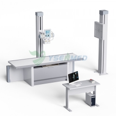
X-ray tables
X-ray room
X-ray generator
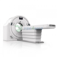
MRI Non-magnetic Equipment
Auto film processors
Manual film processors
X-ray films
X-ray Cassettes
Intensifying screens
Darkroom lights
Film developing solutions
Film dryer
Film viewers
Film hangers
Film markers
Film transfer boxes
Film storing boxes
Chest stands
X-ray dosimeters
X-ray tables
X-ray room
X-ray generator
MRI Non-magnetic Equipment
Hot Products


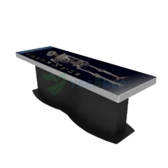

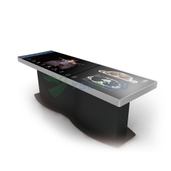


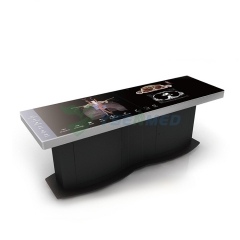


YSDHA-I 88 Digital human virtual anatomy table system
Item No.: YSDHA-I88
| Product parameters | |
| Brand: | YSENMED |
| MOQ: | 1 |
| Package: | Carton Package |
| Ship From: | Guangzhou |
| Lead Time: | 3-5 days |
| Customization: | 7 days |
Description
1Hardware specifications
1.1 Screen: Size: 88-inche( Two 47 inch monitor splices); Resolution: 3840 * 1080; Brightness: 500cd / ㎡ ; Contrast: 1100:1; Touch mode: infrared touch.
1.2 Computer: Processor I7; Graphics card 6G; Memory 16g; Hard disk 500G
1.3 It have at least 3 USB ports and 1 HDMI and 1 VGA for displaying output on the screen.
1.4 The display screen is not less than 2260 x 707 x 750 millimeters (length x width x height),weighs approximately 185 kilograms.
1.5 It has CE and FCC certifications.
2Software specifications
2.1. The system can meet the teaching of systematic anatomy, regional anatomy and sectional anatomy.
2.2 . The system has passed the identification of the Chinese Anatomical Society, and has the software copyright, CE and FCC certification and test report.
2.3. ★ The 3D human body must be reconstructed with the real data of continuous tomography of human body without organic disease and no missing. The data shall be the tomographic data without missing segmental data. The structures such as teeth, male testis, appendix and fat layer must be intact.
2.4. ★ Digital human cross-sectional distance: head and neck are ≤0.5mm, of which the base of the skull is ≤0.1mm, other parts are ≤1.0mm, and the total tomographic data is >2100. Real human body tomographic images with cross-sectional, coronal, and sagittal planes, which can be zoomed in and out at will, with a resolution of ≤0.18mm×0.18mm/pixel.
2.5. The system contains more than 3000 sectional images, including more than 1750 male sectional images, more than 300 male coronal images, more than 500 male sagittal images and more than 490 female sectional images.
2.6. The number of male anatomical 3D structures is more than 2900, and the number of female anatomical 3D structures is more than 260.
2.7. There are more than 1300 anatomical structure labels in the system.
2.8. There are more than 570 image annotation marks in the system.
2.9.★ In the system, the anatomical structure in each fault of the transverse, sagittal and coronal sections shall be circled and marked to facilitate viewing the position and range of each anatomical structure in the fault, and must be associated with the 3D human body. Click any structure position of the 3D or fault, and other areas will respond synchronously.
2.10. Textual description and key structure annotation for anatomical structures, with English name and English pronunciation. After locking the pronunciation status, then click any 3D anatomical structure, it automatically play the English pronunciation .
2.11. The system supports switching the background color of 3D scenes to clearly observe different structures.
2.12. The system supports a variety of operation modes, such as mouse, keyboard and touch screen. Click the structure to view , highlight it immediately, and display notes. Any structure can be zoom in, zoom out, dragged, and observed the front view, back view, side view, upper view, lower view , and observed from any 360 degree angle.
2.13. ★ The directory can display the hierarchy of the structure in the human body, associated dimensions, and their associated dimension marking points.
2.14. The directory structure is a complete system formed by scientifically classifying all the known tissues and organs of the human body from a medical point of view. The directory structure is an index of the entire digital teaching aid.
2.15. All the structures can be browsed through the hierarchical directory and systematic directory, or you can add or delete the organizational structure of a system immediately or select some organizational structures accurately.
2.16. Supporting search, addition, selection and further operation observation of designated human body structure.
2.17. The preset position function is set in the system to facilitate the teacher to establish magnetic stickers according to the teaching content, and quickly find the set 3D human body structure during the teaching. Each preset position magnetic sticker contains the histological slice pictures of the corresponding anatomical structure.
2.18. The structures can be displayed separately, stripped, recovered, dyed, transparent, searched, painted and so on.
2.19. The 3D human body structures can be labeled and described.
2.20. Knowledge expansion: The anatomical structure set in the teaching directory can be used for teaching and learning on the same screen with its associated histological sections, videos and real specimen structures.
2.21. Observation and learning of real specimens: through the anatomical structures in the teaching directory, the real specimens and digital 3D structures associated with them can be compared on the same screen for simultaneous teaching and learning.
3. Systematic anatomy
3.1. There are more than 250 teaching catalogs covering nine systems of human body.As following:Sports system: 45, respiratory system: 15, digestive system: 15, urinary system: 10, vascular system: 50, endocrine system: 5, central nerve: 45, peripheral nerve: 25, reproductive system: 15, visual organ: 10, vestibular cochlear organ: 10, peritoneum:5.
3.2. ★Physiological anatomy of the heart: the beating process of the heart can be observed 360 degrees, the blood flow process of arteries and veins can be displayed, and structures such as ventricles and atria can be separated, valve, papillary muscle, tendon and other structures can be observed under the beating state of the heart.
4. Regional Anatomy
4.1. There are more than 210 regional anatomical parts.As following:Head: 15, neck: 15, chest: 15, abdomen: 35, pelvis and perineum: 20, spine area: 10, upper limb: 50, lower limb: 50.
4.2. The human body structure in the regional anatomy can be stripped layer by layer, and the anatomical incision is marked, and the superficial fascia and deep fascia are kept intact, which is convenient for students to understand the level and adjacent relationship of each part.
4.3. Approach scalpel position: it can display the corresponding anatomical incision of the current local anatomical area, and can be observed in an all-round way.
5. Sectional anatomy
5.1. There are more than 55 human sectional anatomy parts.As following:
Head: 5, neck: 4, chest: 10, abdomen: 10, pelvis and perineum: 5, spine area: 5, upper limb: 10, lower limb: 10.
5.2. There are more than 1700 images of CT/MRI.
5.3. The sectional anatomy includes three parts: real human body section, CT / MR image and 3D structure, and the structures are marked, and the three parts can correspond to each other.
6. Teaching videos
★There are more than 150 teaching videos.As following:Systematic anatomy: 68, regional anatomy: 34, sectional anatomy: 39,nursing:9.
7. Self learning for students
The system is equipped with courseware. The courseware is composed of text, pictures, micro video and 3D anatomical structure. It is an important learning material for students to preview before class and review after class. The courseware also equips with corresponding exercises. The exercises include theoretical exercises and specimen exercises, so as to achieve the purpose of specimen examination. The number of exercises are more than 1800.